|
Beamlines of the Kurchatov specialized source of synchrotron radiation "KISI-Kurchatov"
[an error occurred while processing this directive]
The research activity of synchrotron radiation users is aimed at:
Х nanodiagnostics of ordered organic and bioorganic structures using highly precise spectrally selective X-ray techniques;
Х study of the organic structures and processes in biological objects for genetic engineering, biotechnology, new drugs design;
Х structural diagnostic methods application to ordered nanosystems and multilayer heterocompositions study;
Х study of poly- and single crystals, metal alloys, biological macromolecules solutions; powder diffractometry; X-ray fluorescence elemental analysis;
Х convergent visualization approaches application to cultural heritage samples;
Х scientific basis creation in the field of combined complementary synchrotron-neutron research;
Х X-ray absorption spectroscopy methods EXAFS / XANES development;
Х new medical diagnostics methods development.
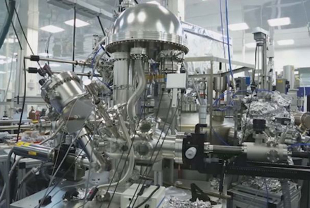
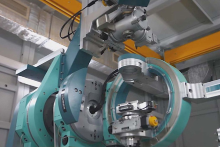
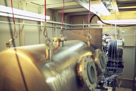
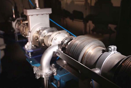
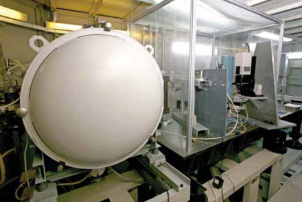
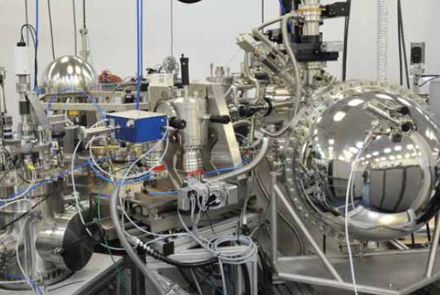
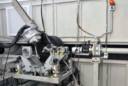
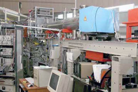
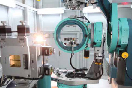
"KISI-Kurchatov" is equipped with a set of beamlines, including monochromatization and intense radiation beam control units, vacuum chambers, precision goniometric equipment, X-ray optical elements, detectors, automation and experiment control systems, etc.
LANGMUIR
Beamline for x-ray studies of molecular films on liquid surfaces. The main feature of the beamline is the X-ray optical scheme, which provide possibilities of SR beam deviation from horizontal plane for X-ray measurements on the liquid.
Methods:
Х X-ray reflectometry (XRR) - electron density profile along the depth of the structure - thickness, density, roughness of the layers;
Х X-ray Standing waves (XSW) - ion distribution profile over the depth of the structure;
Х Grazing incidence diffraction (GID) - the crystal structure of two-dimensional systems (lattice parameters, molecular structure, molecular tilt angles, correlation lengths);
Х Total X-ray Reflection Fluorescence (TXRF) - the chemical composition of the surface layers.
BIOSAX
Beamline for studying biological objects structure in statics and dynamics by diffraction and scattering methods in small and wide angles region (SAXS / WAXS). The main researches are aimed at determining the structure and structural dynamics of various types of tissues of animals and humans and soft matter objects.
Methods:
Х Small-angle X-ray scattering (SAXS) - a study of the structural parameters of samples with characteristic ordering sizes in the range 0.2Ц200 nm, with the ability to change the temperature of the sample from -50 C to +150 C;
Х X-ray powder diffractometry (XRD) - determination of the phase composition, crystallite sizes, texture, microstresses.
RSA (XRD)
Beamline for studying atomic and real structures using single- and polycrystalline methods using 2D detector.
Methods:
Х X-ray diffraction analysis of single-crystal inorganic objects. Spatial structure with atomic resolution;
Х Precision powder diffraction with a 2D scanning detector. Phase composition, crystallite sizes, texture, microstresses.
PHASE
Specialized beamline for diagnostics using methods of precision X-ray diffractometry and reflectometry, as well as phase-sensitive methods for studying substances.
Methods:
Х X-ray Standing waves (XSW);
Х High-resolution diffraction (XRD);
Х Multiwave diffraction;
Х Surface diffraction;
Х X-ray acoustooptics;
Х X-ray holography;
Х Resonance diffraction;
Х X-ray Reflectometry (XRR);
Х Diffuse scattering.
BELOK (PROTEIN)
Protein crystallography beamline for X-ray diffractometry of macromolecular single crystals in a wide range of radiation energies. Resolution is 1Ц1.2 ≈ for crystals with lattice parameters up to 100≈, and a resolution of 1.6Ц1.8≈ for crystals with lattice parameters of 200≈.
Methods:
Х X-ray diffraction analysis of macromolecular crystals with large cell parameters;
Х Monocrystalline diffractometry - determination of the spatial structure;
Х Powder diffractometry.
RKFM
X-ray crystallography and physical material science beamline for materials structure characterization by X-ray diffraction and scattering methods.
Methods:
Х Double and triple axis high resolution diffractometry (XRD, HRXRD);
Х Multiwave diffraction;
Х X-ray topography;
Х Reciprocal space mapping (RSM);
Х X-ray Reflectometry (XRR);
Х X-ray standing waves (XSW);
Х X-ray fluorescence analysis (XRF);
Х X-ray acoustooptics.
PRO
Beamline of precision X-ray optics, focused on x-ray diffraction methods for the study of matter.
Methods:
Х Double and triple axis high resolution diffractometry (XRD, HRXRD);
Х Multiwave diffraction;
Х X-ray Reflectometry (XRR);
Х X-ray standing waves (XSW);
Х Resonance diffraction.
STM
Structural material science beamline for studying the features of the spatial structure of functional materials in a wide scales range.
Methods:
Х EXAFS / XANES - chemical state of the absorbing atom and its local environment;
Х X-ray powder diffraction (XRD) Ч crystalline structure;
Х Small-angle scattering (SAXS) - sizes and shape of nanoparticles.
REFRA
This beamline uses refractive optics, designed to study materials by methods of EXAFS spectroscopy in the fluorescence mode and X-ray fluorescence elemental analysis, and also allows samples mapping.
EXAFS-D
Beamline for polycrystalline and amorphous materials study by X-ray spectroscopy and diffraction.
Methods:
Х Diffraction in the geometry of forward and backscattering; high sensitivity to deformations and microstresses;
Х Energy dispersive EXAFS with millisecond resolution (chemical state of the absorbing atom and its local environment).
NANOPES
Beamline for studying the electronic structure of solids by photoelectron, optical and probe spectroscopy. It operates in ultrahigh vacuum, the energy range of photon energies from vacuum ultraviolet to soft X-ray (25-1500 eV).
Methods:
Х Angular resolution photoelectron spectroscopy (ARPES):
Х Depth scanning;
Х Near Edge X-ray absorption (NEXAFS);
Х Subtle features of the PE spectra of the valence band;
Х Study of deep levels;
Х SPM (STM + AFM) microscopy.
RT-MT
X-ray topography and microtomography beamline. Studying of samples with typical sizes from 2 to 50 mm with inhomogeneities in 2 - 1000 мm range, respectively.
Methods:
Х X-ray microtomography;
Х X-ray topography.
MEDIANA
Beamline is intended for medical and materials science diagnostics. Beamline consists of two units assembled on one channel - a high-pressure diffractometer and an X-ray introscopy unit.
Methods:
Х Absorption introscopy;
Х Phase-contrast introscopy;
Х Tomography - obtaining images of the internal structure of objects, incl. for biomedical purposes;
Х Diffractometry at high pressures - the structure of polycrystalline objects.
LIGA
Currently used to obtain three-dimensional images of objects ranging in size from 1 to 10 cm using the method of X-ray computed tomography. Beamline is focused on research in biology, materials science, paleontology, etc.
|

